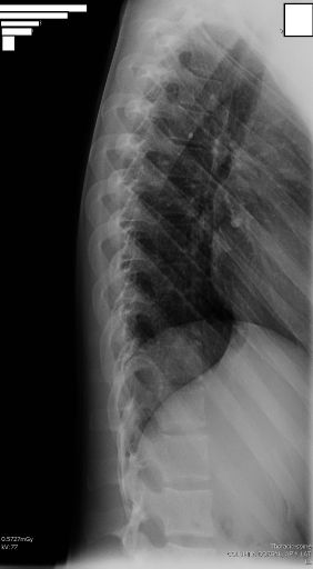
LAT THORACIC
Lateral Thoracic Spine Projection • Evaluation of vertebral bodies and intervertebral discs
Visible Anatomical Structure
The following must be clearly observed:
- Thoracic vertebral bodies (T1 to T12)
- First and second lumbar vertebrae (L1 and L2)
- Posterior part of the ribs
- Intervertebral foramina (neural foramina)
- Intervertebral spaces (discs)
- Spinous processes in superimposition
- Pedicles in lateral view
Plate Size and Orientation
Longitudinal orientation to cover the entire thoracic spine and upper lumbar vertebrae
Choice of Side: Left Preferred
"Standard protocol is left lateral decubitus"
Reasons for preferring the left side:
- Decreases cardiac silhouette in the image
- Better visualization of vertebral bodies
- Reduced superimposition with mediastinal structures
- Standardized protocol for serial comparisons
Main advantage: The heart projects anteriorly, reducing superimposition with the thoracic spine
Patient Positioning
Lateral Decubitus (Preferred Position)
Alternative Position: Upright (Erect)
When lateral decubitus is not possible:
- Perform standing at the upright bucky
- Same alignment and centering criteria
- Useful for patients with pain or difficulty lying down
Lumbar Support Details
"Support must be placed under the lumbar area" so that:
- The longitudinal axis of the spine is parallel to the table plane
- Maintain alignment between ankles and table
- Maintain alignment between the ankles
- Maintain alignment between the knees
- Maintain alignment between knees and table
Purpose: Prevent torso rotation and maintain the spine in a true lateral position
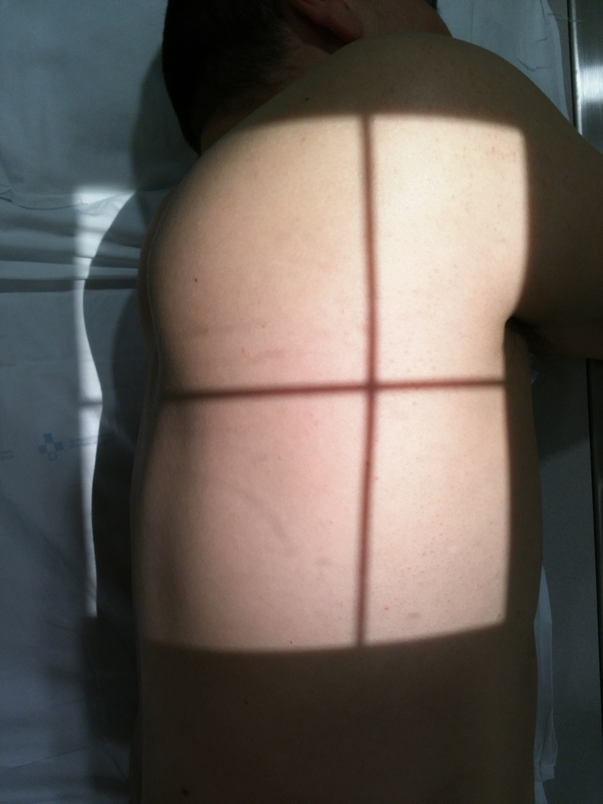
Central Ray Point
Location: Sixth thoracic vertebra
Lateral Decubitus: Vertical ray perpendicular to the cassette
Upright: Horizontal ray perpendicular to the cassette
Goal: Centered at the midpoint of the thoracic spine
Optimal Image Characteristics
Vertebrae T1-T12
All included in the field
Intervertebral Discs
Well-defined spaces
Intervertebral Foramina
Neural foramina visible
Posterior Ribs
Projected without critical superimposition
Alignment
Physiological curvature preserved
Transitions
L1 and L2 visible
Common Technical Challenges
Frequent issues in lateral thoracic projection:
- Patient rotation leading to an oblique image (lack of lumbar support)
- Rib superimposition over vertebral bodies
- Cardiac silhouette obscuring middle thoracic vertebrae (right side)
- Breathing during exposure causing blurriness
- Motion due to lack of leg flexion
- Incomplete field not including T1 or L1/L2
- Marked kyphosis making full visualization difficult
Solution: Use left lateral decubitus, adequate lumbar support, leg flexion, and instruct breath-hold
Recommended Breathing Technique
To reduce respiratory motion:
- Instruct the patient to breathe in deeply
- Exhale completely before exposure
- Hold breath (apnea) during exposure
- Do not move during image acquisition
Special Considerations
Geriatric Patients
Marked kyphosis may require multiple exposures or a double-cassette technique.
Obese Patients
Increase kV and mAs; consider a non-grid technique for very obese patients.
Patients with Pain
An upright position may be better tolerated than lateral decubitus.
Severe Kyphosis
Consider AP projection instead of lateral for initial evaluation.
Patient Instructions
Specific instructions for lateral decubitus:
1. "Lie down on your left side"
2. "Flex your knees to be comfortable"
3. "Place your elbows in front, flexed at 90°"
4. "Take a deep breath and then let it all out"
5. "Hold your breath and do not move"
6. "Stay still until I tell you"